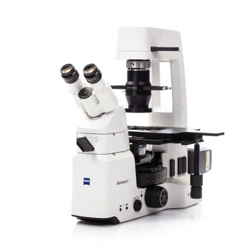
An Inverted microscope unique feature is that the objective lens is positioned below the specimen, while the light source and condenser are above. Its light source and condenser position above the stage, directing light downward, while the objectives and turret are situated below the stage, pointing upward, versus an Upright microscope is a type of microscope where the light source, condenser, and objectives are positioned above the stage, while the stage itself is located beneath them. This configuration allows the light to pass through the specimen from below and enables observation from the top of the microscope.
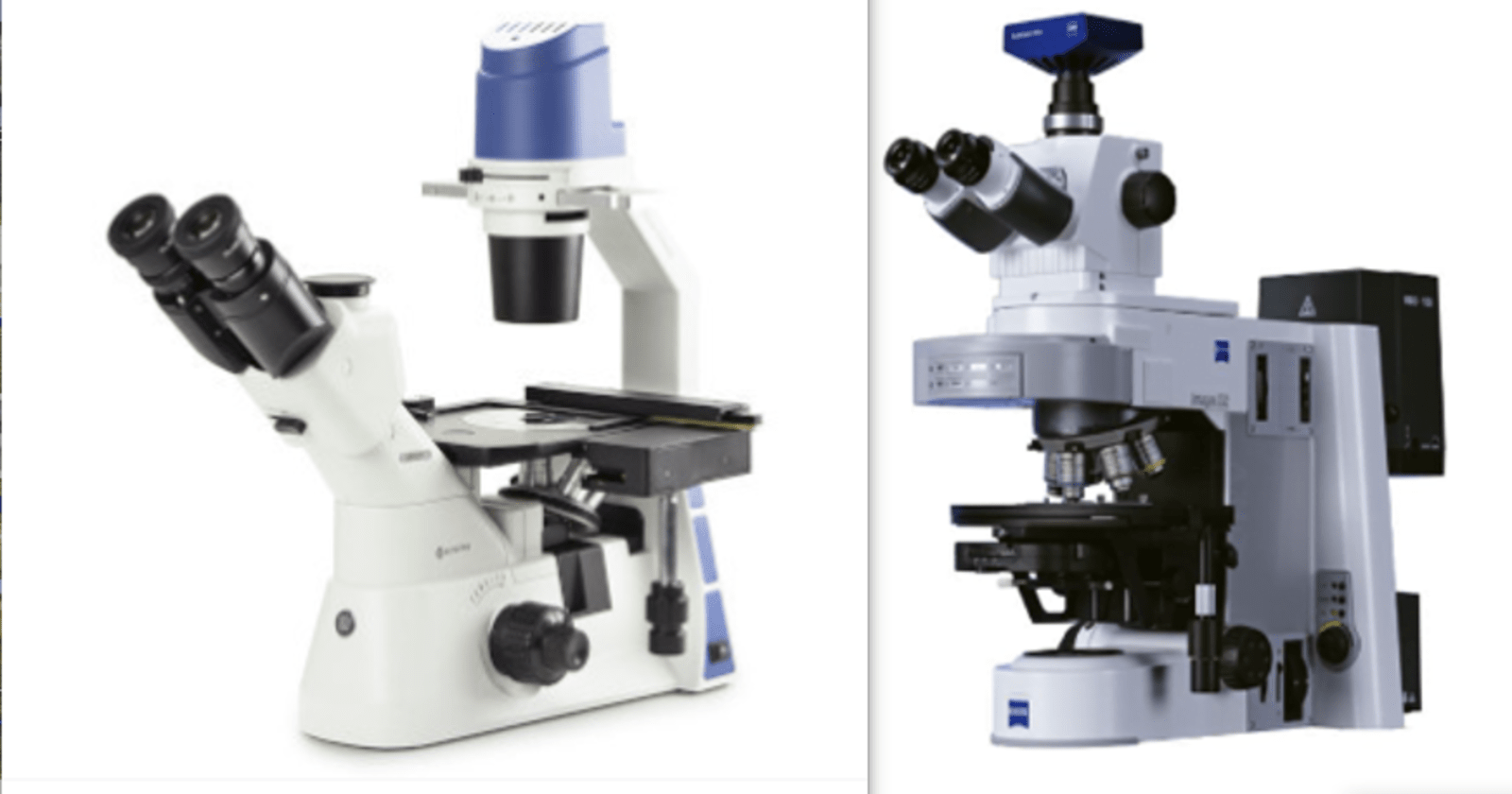
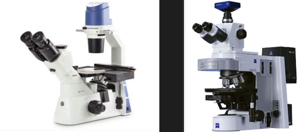
Inverted Microscope vs Upright Microscope
Inverted Microscope
An inverted microscope is characterized by having its light source and condenser positioned above the stage, directing light downwards, while the objectives and turret are located below the stage, facing upward.
In an Inverted microscope, the objective lens is below the stage where the sample is placed. You view the sample from underneath. To make it similar to using an upright microscope, the optical path is angled upwards towards the observation tube and eyepieces.
The Inverted microscope’sdesign is tailored to the unique needs of cell biology and live-cell imaging, offering advantages for observing specimens in culture containers and providing an ergonomic viewing angle for researchers.
This design allows for various applications, especially those involving live-cell imaging and cell culture work.
An upright microscope is a type of optical microscope that is designed for observing samples in an upright position, meaning that the specimen is placed on a stage and viewed from above.
This microscope is commonly used in laboratories, schools, and research settings for magnifying and studying small objects such as cells, tissues, or microorganisms. It typically consists of a light source, lenses, and an eyepiece, allowing users to view and analyze the details of the specimen at various magnifications. The term “upright” distinguishes it from an inverted microscope, where the specimen is viewed from below.
4 Important features of orientation of the optical components of an Inverted Microscope vs an upright microscope
Inverted microscope
Upright microscope
light is directed from above the sample through its optical component
The optical components align for a straight light path from the objective lens to the eyepiece.
The sample is typically placed on a specialized stage above the objective lens.
The sample is usually placed on a slide or a stage below the objective lens.
Objective lens, positioned above the stage and facing downward, magnifies the image.
Objective lens, below the stage, faces upwards toward the specimen, magnifying the image.
Observers look up through the eyepiece to view the specimen.
Observers look down through the eyepiece to view the specimen.
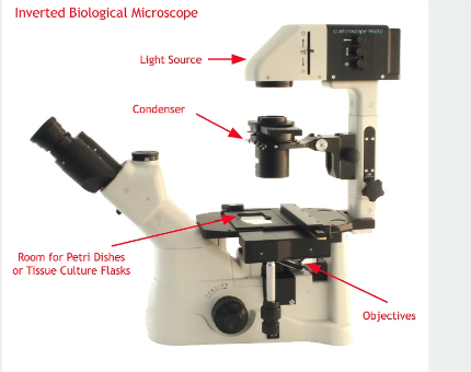
Basic Components of an Inverted Microscope vs an Upright Microscope
showing the basic components of an Inverted microscope
Inverted microscopes and upright microscopes are both essential tools used in microscopy, but they differ in their designs and applications.
Upright Microscope
Inverted Microscope
Eyepiece (Ocular) Located at the top of the microscope.
Eyepiece (Ocular) Located at the bottom of the microscope.
Objective Lenses Mounted on a rotating nosepiece below the eyepiece.
Objective Lenses Also mounted on a rotating nosepiece, but above the stage.
Nosepiece Holds and allows rotation of the objective lenses.
Nosepiece Holds and allows rotation of the objective lenses.
Stage Located below the objective lenses.
Stage Located above the objective lenses.
Condenser Beneath the stage, focuses light onto the specimen.
Condenser May be present, but not as prominent as in upright microscopes.
Illuminator Typically at the base of the microscope.
Illuminator Usually located above the stage.
Diaphragm Adjusts the amount of light reaching the specimen.
Diaphragm May be present, similar to an upright microscope.
Coarse and Fine Focus Adjustment Controls for initial and precise focusing.
Coarse and Fine Focus Adjustment Controls for initial and precise focusing.
Additional Features – Mechanical stage with controls.
Additional Features – Larger stage for accommodating heavy specimens.
– Often used for routine microscopy.
– Commonly used in cell culture work.
– Well-suited for various specimens.
– Often equipped with a heated stage for live cell imaging.
5 Essential ways in which an inverted microscopes are best used
Cell Culture Work:
Observation of Living Cells: Inverted microscopes are ideal for observing cells cultured in dishes or flasks. The positioning of the objective lens beneath the sample allows for easy access and manipulation of cells without the need to disturb the culture vessel.
Monitoring Cell Growth and Behavior: Researchers can use inverted microscopes to monitor the growth, proliferation, and behavior of cells over time. This is crucial in cell culture studies, where maintaining optimal conditions for cell health is essential.
Cellular Interactions: Inverted microscopes are employed to study cell-cell interactions and the effects of various treatments on cultured cells. This is particularly important in
Time-Lapse Imaging: Inverted microscopes are well-suited for time-lapse imaging, allowing researchers to capture and analyze dynamic processes occurring within living cells over an extended period. This is beneficial for studying phenomena like cell division, migration, and response to stimuli.
Fluorescence Microscopy: Many inverted microscopes are equipped with fluorescence capabilities, enabling the visualization of specific cellular structures or molecular markers tagged with fluorescent dyes. This is widely used in molecular biology and biomedical research.
Confocal Microscopy: Some advanced inverted microscopes are compatible with confocal microscopy techniques. Confocal microscopy provides enhanced optical sectioning, reducing out-of-focus blur and allowing for three-dimensional imaging of cellular structures.
3. Microinjection and Micromanipulation:
Inverted microscopes are often used for microinjection and micromanipulation techniques. Researchers can precisely inject substances into individual cells or manipulate cellular structures using specialized micromanipulation tools.
4. Electrophysiology Studies:
In neuroscience and related fields, inverted microscopes are employed for electrophysiology studies. Researchers can use these microscopes to visualize and manipulate neurons or other excitable cells during electrophysiological experiments.
5. IVF (In Vitro Fertilization) Procedures:
In the field of reproductive medicine, inverted microscope are used for examining oocytes and embryos during IVF procedures. The inverted design facilitates easy access and manipulation of gametes and embryos in culture dishes.
9 Essential ways in which upright microscopes are best used
Biological Research:
Cellular and Tissue Examination: Upright microscopes are essential tools in biology for studying the structure and function of cells and tissues. They enable researchers to observe details such as cell morphology, organelles, and cellular processes.
Microorganism Studies: Microbiologists use upright microscopes to study bacteria, fungi, and other microorganisms. This is crucial for understanding microbial structures, behaviors, and interactions.
Clinical Diagnostics:
Medical Pathology: In medical laboratories, pathologists use upright microscopes to examine tissue samples for diagnosing diseases. This includes the identification of cancerous cells, infections, and abnormalities.
Hematology: Hematologists use microscopes to analyze blood samples, identifying blood cells, abnormalities, and disorders.
Material Science:
Material Analysis: Upright microscopes are used in material science for examining the microstructure of materials. This is vital for understanding material properties, defects, and composition.
Quality Control: Industries use upright microscopes for quality control processes, ensuring that materials and products meet specific standards and specifications.
Education:
Teaching and Learning: Upright microscopes play a crucial role in educational settings, allowing students to explore the microscopic world. They are commonly used in classrooms and laboratories for teaching biology, anatomy, and various scientific disciplines.
Forensic Science:
Forensic Analysis: Forensic scientists use microscopes to analyze evidence such as hair, fibers, and biological samples. This aids in criminal investigations and the resolution of legal cases.
Botany and Plant Science:
Plant Anatomy: Botanists use upright microscopes to study plant structures, including tissues, cells, and reproductive organs. This helps in understanding plant development and physiology.
Entomology:
Insect Studies: Entomologists use microscopes to examine the anatomy and morphology of insects. This is important for species identification, understanding behaviors, and studying the ecological role of insects.
Fluorescent Labeling: Upright microscopes are often used in fluorescence microscopy, where fluorophores are used to label specific structures or molecules. This technique enhances contrast and allows for the visualization of specific components within cells.
Photomicrography:
Image Documentation: Upright microscopes can be used for photomicrography, capturing high-quality images of microscopic specimens. These images are important for documentation, analysis, and presentation of research findings.
Ways in which upright microscopes vs inverted microscope are best used
Upright Microscopes
Inverted Microscopes
Sample Type Typically used for viewing solid or opaque samples placed on slides.
Sample Type Ideal for observing living cells and specimens in culture dishes or containers.
Working Distance Short working distance, limiting space between the objective lens and the sample.
Working Distance Longer working distance, providing more space for manipulation and intervention.
Sample Manipulation Limited ability for manipulation due to the positioning of the stage.
Sample Manipulation Convenient for manipulating and accessing samples from the top, making it suitable for micromanipulation and microinjection.
Observing Thick Samples Suitable for thin samples on slides; may require sample sectioning for thicker specimens.
Observing Thick Samples Well-suited for thick or 3D samples without the need for extensive sample preparation.
Light Source Light is typically transmitted from below the sample through the condenser.
Light Source Light is often directed onto the sample from above, which is advantageous for certain techniques like phase contrast and DIC.
Live Cell Imaging Less convenient for observing live cells due to limitations in manipulating culture vessels.
Live Cell Imaging Excellent for observing live cells in culture, as the objectives can focus through the bottom of culture dishes.
Microscopy Techniques Commonly used for brightfield, darkfield, and fluorescence microscopy.
Microscopy Techniques Widely used for phase contrast, DIC, and fluorescence microscopy, particularly for live-cell imaging.
Applications Commonly used in histology, pathology, and routine laboratory work.
Applications Preferred for applications such as cell culture, in vitro fertilization, and time-lapse imaging of living cells.
Cost Generally more affordable compared to inverted microscopes.
Cost Inverted microscopes tend to be more expensive due to their specialized design and features.
Educational Use Often used in educational settings for teaching basic microscopy techniques.
Educational Use Suitable for advanced microscopy techniques, making it valuable in research and advanced educational settings.
It’s important to note that the choice between upright and inverted microscopes depends on the specific requirements of the microscopy application and the nature of the samples being studied. Each type of microscope has its own advantages and is better suited to certain types of observations.
Advantages of an Inverted microscope vs Advantages of an Upright microscope
Live Cell Imaging: Inverted microscopes are ideal for observing living cells in culture. The design allows for easy manipulation and observation of cells in containers like petri dishes and culture flasks.
Manipulation and Intervention: The inverted configuration provides a larger working space between the objective lens and the sample, making it easier to manipulate and intervene with the specimen. This is particularly advantageous for procedures like micromanipulation and microinjection.
Thick and 3D Sample Observation: Inverted microscopes are well-suited for observing thick or 3D samples without the need for extensive sample preparation. The longer working distance allows for the examination of specimens with varying depths.
Phase Contrast and DIC Imaging: Inverted microscopes are commonly equipped with phase contrast and differential interference contrast (DIC) optics, making them suitable for detailed imaging of transparent or low-contrast specimens.
Non-Destructive Observation: The design allows for non-destructive observation of samples in their natural environment, making it suitable for long-term time-lapse imaging studies.
Sample Accessibility: The sample is easily accessible from the top, allowing for easy addition of reagents, fluids, or other materials during the observation process.
Specialized Techniques: Inverted microscopes are well-suited for specialized techniques like fluorescence microscopy, making them valuable in various fields such as cell biology and molecular biology.
Cell Culture Applications: In research and medical laboratories, inverted microscopes are commonly used for observing and working with cells in culture, making them essential for applications like in vitro fertilization.
Contamination Prevention: The design ensures that the specimen remains uncontaminated by avoiding direct contact with the objective lens.
Sterility Maintenance: This feature contributes to maintaining the sterility of the sample during observation, making it suitable for applications where sample purity is crucial.
Cost: Upright microscopes are generally more cost-effective than inverted microscopes. This makes them more accessible for educational institutions and routine laboratory work.
Educational Use: Upright microscopes are commonly used in educational settings for teaching basic microscopy techniques. They are straightforward to use and provide a simple platform for learning microscopy principles.
Histology and Pathology: In routine laboratory work, upright microscopes are often used for histology and pathology applications. They are suitable for examining thin sections of tissues on glass slides.
Brightfield and Darkfield Imaging: Upright microscopes are well-suited for brightfield and darkfield microscopy techniques. They are versatile for a range of routine observations in various scientific disciplines.
Simple Sample Preparation: Sample preparation for upright microscopes is often simpler, as thin sections on slides are the common format. This can be advantageous for quick examination of prepared samples.
Versatility: Upright microscopes are versatile and suitable for various microscopy techniques, including basic fluorescence microscopy. They can cover a broad range of applications in research and routine laboratory work.
Routine Laboratory Applications: In fields such as microbiology, hematology, and general biology, upright microscopes find applications in routine laboratory analyses.
Stability and Durability: Upright microscopes are generally more stable due to their design. This stability can be advantageous for routine observations that do not require constant manipulation of the sample.
Disadvantages and limitations of an Inverted microscope
Cost: Inverted microscopes can be more expensive than traditional upright microscopes. The complexity of their design, including the need for special objectives and condensers, can contribute to higher costs.
Objective lenses: Specialized objectives are required for inverted microscopes due to their unique design. These objectives can be more expensive than those used in upright microscopes.
Limited working distance: Inverted microscopes often have a limited working distance, which is the distance between the objective lens and the specimen. This limitation can be a challenge when working with large or three-dimensional specimens.
Sample size and thickness: Inverted microscopes may have restrictions on the size and thickness of samples that can be accommodated. This can be a limitation when working with certain types of samples, especially those that require specific sample preparation techniques.
Accessibility and manipulation: Accessing and manipulating specimens can be more challenging with an inverted microscope, especially when compared to upright microscopes. It may be difficult to manipulate samples or perform interventions during live imaging.
Image quality: While inverted microscopes are suitable for many applications, their design may result in slightly lower image quality compared to some specialized upright microscopes. This is particularly relevant for certain imaging techniques and high-resolution applications.
Limited versatility: Inverted microscopes are specifically designed for certain applications, such as cell culture and live cell imaging. They may not be as versatile as upright microscopes for other types of microscopy, such as stereomicroscopy or certain advanced imaging techniques.
Vibration sensitivity: Inverted microscopes can be more sensitive to vibrations because of their design. This sensitivity can impact image quality, especially when conducting high-magnification imaging or delicate manipulations.
Objective immersion limitations: Immersion objectives, commonly used for high-resolution imaging, may be more challenging to use with inverted microscopes. The need for immersing the objective in a liquid medium can complicate specimen handling and manipulation.
Disadvantages and limitations of an Upright microscope
Limited Depth of Field:
Upright microscopes typically have a limited depth of field, making it challenging to focus on different layers of thick specimens simultaneously. This can be a drawback when studying three-dimensional structures.
Limited Magnification:
While upright microscopes can achieve high magnifications, there is a practical limit to how much magnification they can provide. For extremely high magnifications, other types of microscopes, such as electron microscopes, may be more suitable.
Limited Field of View:
At high magnifications, the field of view may become significantly smaller, making it difficult to observe larger specimens or get an overview of a sample.
Inability to Observe Living Organisms in Natural Conditions:
Upright microscopes are often used with prepared slides, which may involve fixing and staining specimens. This process can alter the natural state of living organisms, making it challenging to observe them in their native environment.
Not Suitable for Thick or Opaque Samples:
Upright microscopes may struggle with specimens that are too thick or opaque for light to pass through effectively. In such cases, other imaging techniques or microscopy methods may be more appropriate.
Artifact Formation:
Sample preparation methods, such as staining, can introduce artifacts that may affect the accuracy of observations. Additionally, the interaction of light with certain materials may lead to optical artifacts.
Limited Resolution:
The resolution of an upright microscope is limited by the wavelength of light. This limitation can make it challenging to distinguish very small details in specimens.
Require Adequate Lighting Conditions:
Upright microscopes rely on external light sources, and variations in lighting conditions can affect the quality of observations. Proper illumination is crucial for obtaining clear and accurate images.
Maintenance and Alignment:
Upright microscopes require regular maintenance and alignment to ensure optimal performance. If not properly maintained, issues such as misalignment can lead to decreased image quality.
Cost:
High-quality upright microscopes with advanced features can be expensive, which may be a limitation for some laboratories or researchers with budget constraints
key physical features when identifying an Inverted microscope vs Upright microscope
1. Objective Lens Placement:
Upright Microscope: The objective lenses are positioned above the specimen, and light passes through the specimen from below.
Inverted Microscope: The objective lenses are located below the specimen stage, and light passes through the specimen from above.
2. Stage Orientation:
Upright Microscope: The stage is typically located below the objective lenses, and the specimen is placed on top of the stage.
Inverted Microscope: The stage is situated above the objective lenses, and the specimen is mounted on the bottom of the stage or in special containers.
3. Light Source Location:
Upright Microscope: The light source is usually positioned beneath the stage, illuminating the specimen from below.
Inverted Microscope: The light source is typically above the stage, directing light downward onto the specimen.
4. Specimen Handling:
Upright Microscope: Specimens are often placed on slides and coverslips on top of the stage.
Inverted Microscope: Specimens can be cultured in containers such as Petri dishes or flasks, and the microscope objectives reach up to view them from below.
5. Applications:
Upright Microscope: Commonly used for routine laboratory work, histology, and examining thin sections of biological samples on slides.
Inverted Microscope: Preferred for applications like cell culture, live-cell imaging, and observations of larger and three-dimensional specimens.
6. Focus Adjustment:
Upright Microscope: The focus adjustment knobs are typically on the side or at the back of the microscope.
Inverted Microscope: The focus adjustment knobs are often on the front or at the top of the microscope, as they are accessible when viewing specimens from below.
These features highlight the main differences between inverted and upright microscopes. The choice between them depends on the specific needs of the microscopy application, such as the type of specimens being observed and the imaging techniques employed.


 vs Pumpkin")


 vs Pumpkin")

