O-Arm vs C-arm: for surgeries,Radiation exposure,differences,advantages
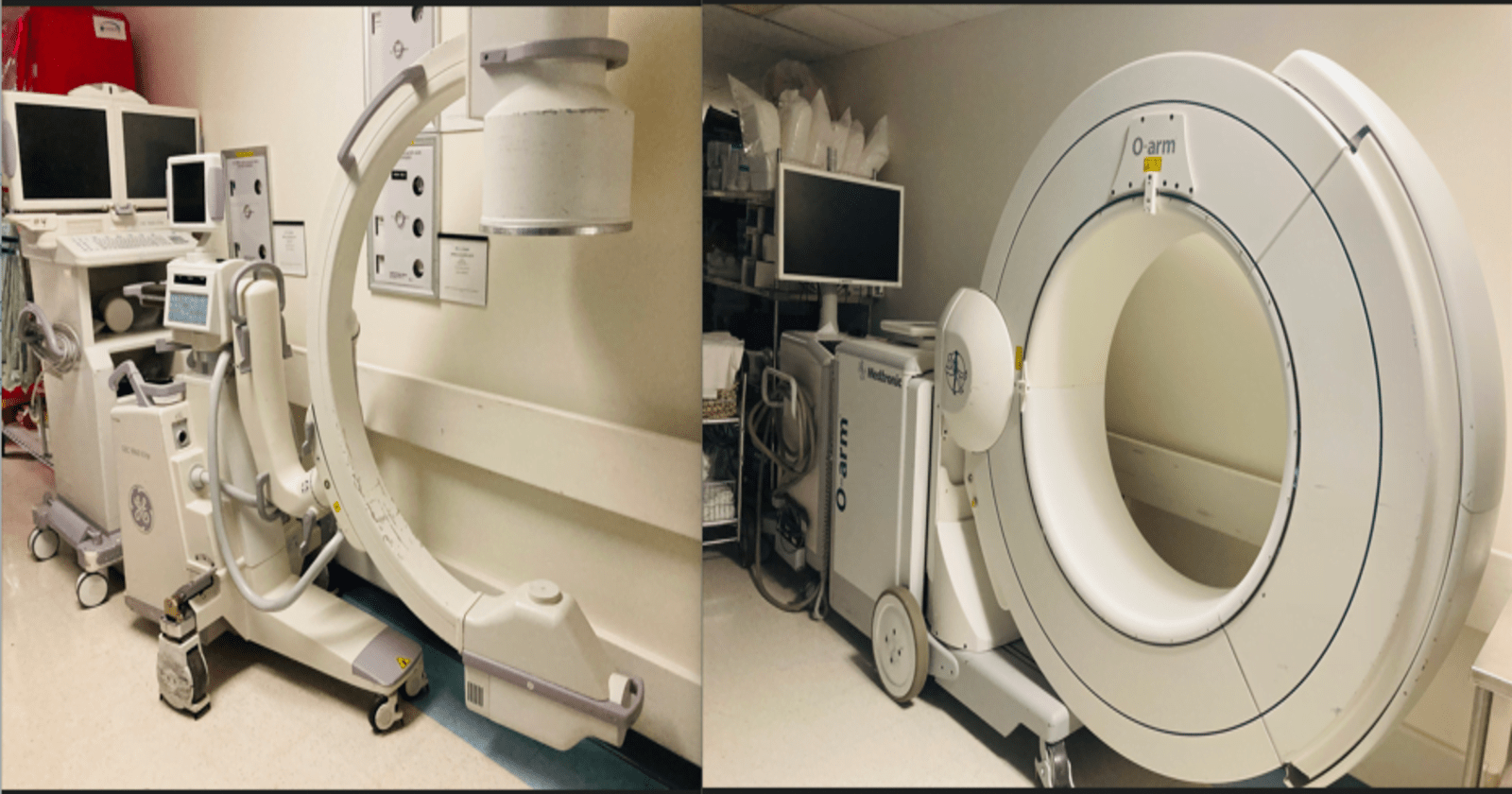
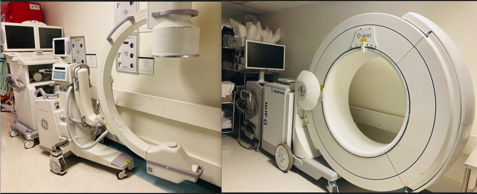
A C-arm is a medical imaging device with a C shape that provides fluoroscopic X-ray images in real-time versus an O-Arm which is a type of intraoperative 3D imaging device with an O shape, that provides real-time, high-quality, 3D images of the patient’s anatomy during surgery.
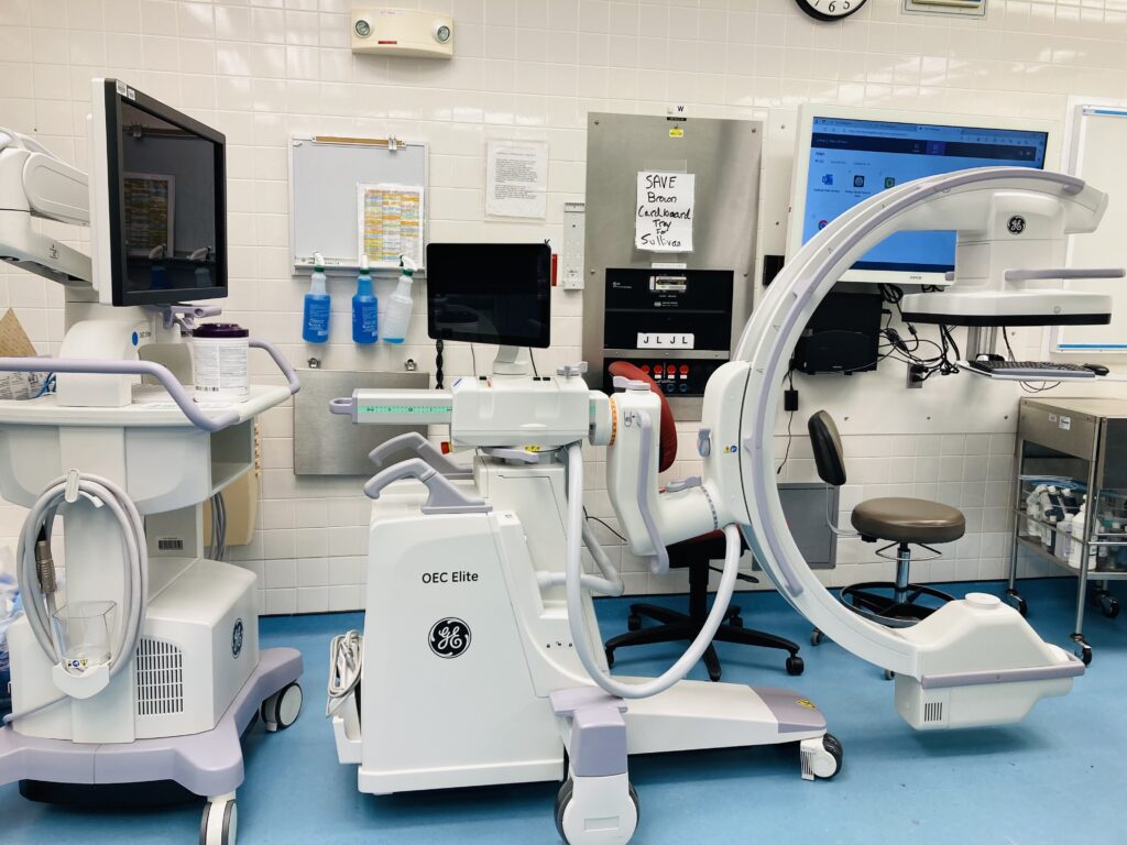
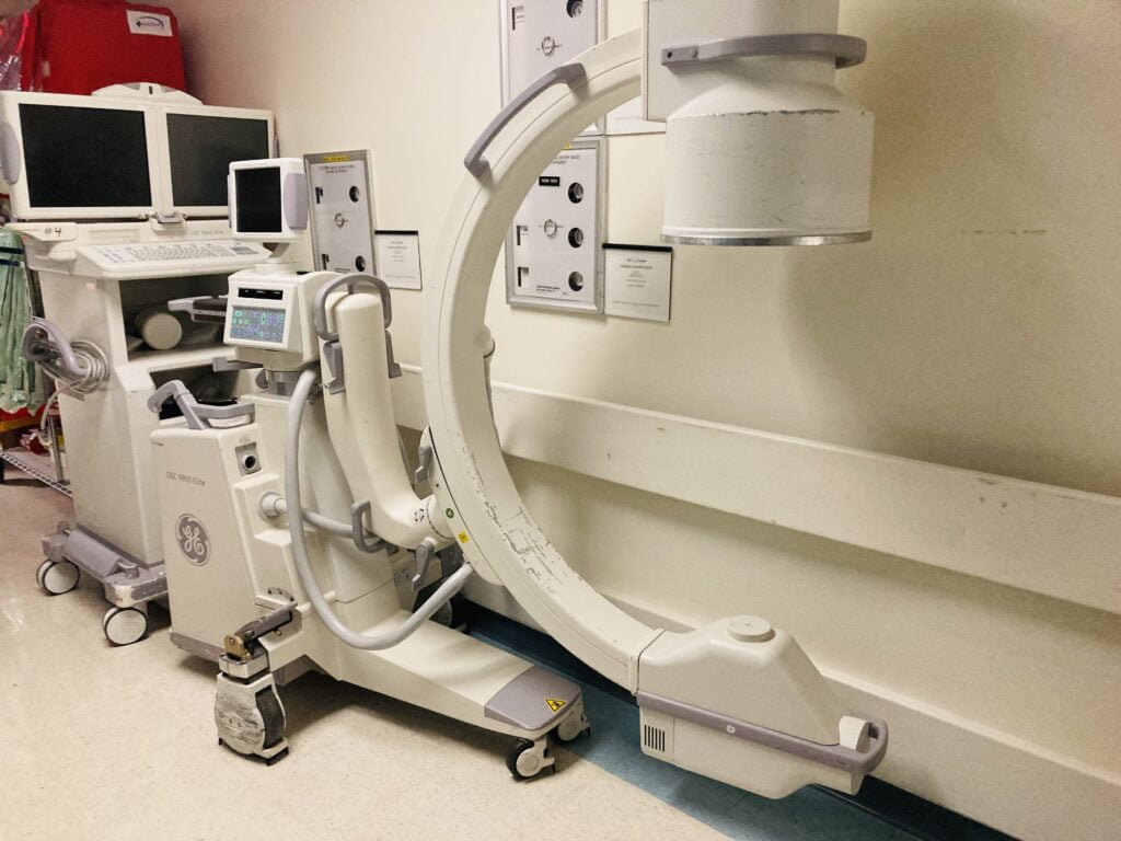
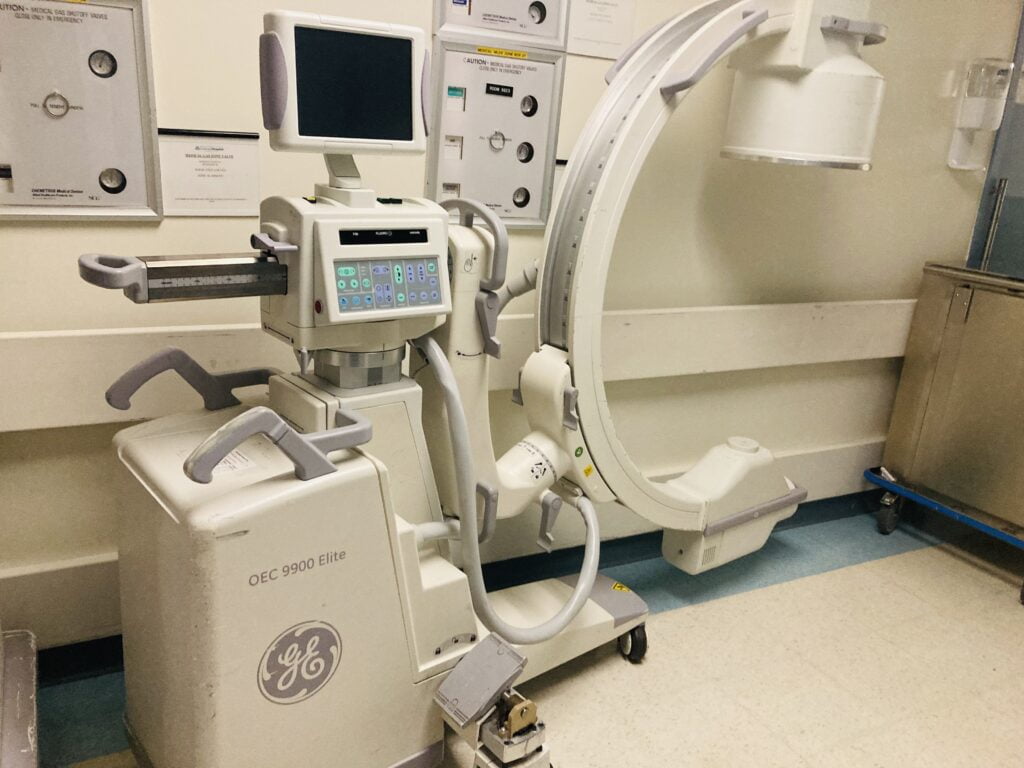
C-arm imaging device
C-arms are more general-purpose more Versatile, mobile, and widely used for various procedures that require real-time X-ray imaging. while O-arms are specialized for high-quality 3D imaging in specific surgical applications, particularly beneficial for complex procedures requiring detailed anatomical visualization, such as spinal surgeries and certain neurosurgical interventions.
The name “C-arm” comes from the C-shaped design of the device, which consists of a curved, arm-like structure that can be easily maneuvered and positioned around a patient.
The C-arm is widely used in various medical procedures, especially in surgical and interventional settings, to provide dynamic imaging during the course of a procedure.
C-Arm imaging device vs O-Arm imaging device
The O-arm system consists of a O-arm (a large, arc-shaped imaging device) that can be positioned around the patient during surgery to capture 2D and 3D images. Surgeons can use these images to enhance their understanding of the patient’s anatomy, guide the placement of implants, and verify the success of the procedure in real time.
Various surgical procedures where real-time imaging guidance is needed.
Less flexible due to fixed installation.
Highly flexible, can be easily positioned for different angles.
Commonly used in spinal and neurosurgical interventions.
Suitable for a wide range of procedures, including orthopedic, vascular, and cardiac surgeries.
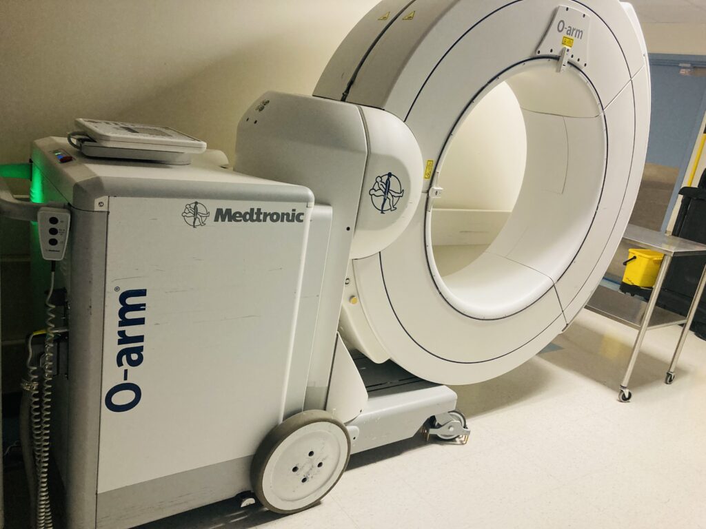
Understanding the O-Arm and what it dose
The O-arm is a state-of-the-art intraoperative imaging system designed to provide real-time, high-quality, three-dimensional images of a patient’s anatomy during surgery. It is particularly used in orthopedic and spinal procedures, where precise visualization of the surgical site is critical for accurate placement of implants and instruments.
The O-arm employs a combination of fluoroscopy and 3D imaging technology, allowing surgeons to have a comprehensive view of the patient’s anatomy in various planes, including axial, sagittal, and coronal.
This level of detailed imaging aids in achieving greater accuracy and safety in surgical procedures, reducing the risk of complications and improving patient outcomes.
One of the distinctive features of the O-arm is its ability to capture high-resolution images in real time, enabling surgeons to make immediate, informed decisions during surgery. This real-time feedback loop enhances surgical precision and allows for adjustments as needed, ultimately leading to more successful procedures.
Overall, the O-arm represents a significant advancement in intraoperative imaging, revolutionizing the way surgeons approach complex orthopedic and spinal surgeries, and contributing to improved patient care.
O-ARM Used in the operating room
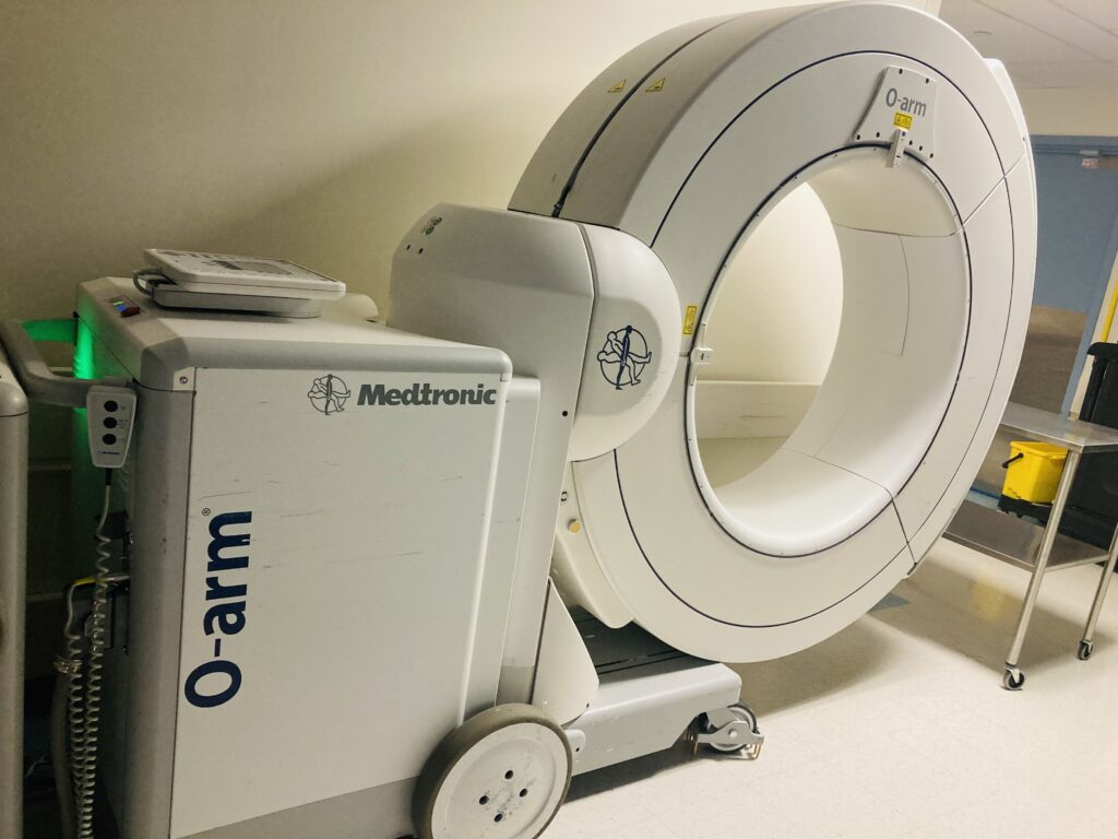
The O-arm surgical imaging system was first introduced in 2007 by Medtronic, a medical technology company. The O-arm provides high-quality, real-time, 2D and 3D imaging during surgical procedures, particularly in the field of orthopedics and neurosurgery. It has since become an important tool in the operating room, allowing surgeons to have precise and detailed imaging guidance during complex procedures
Radiation Exposure Level of the O-Arm vs the C-Arm
O-arm
C-arm
Low
Moderate to High
3D imaging with low dose
2D imaging with higher dose
Precise radiation control with reduced scatter
Less precise radiation control with potential scatter
Commonly used in spine and orthopedic surgeries
Widely used in various procedures
Radiation dosage typically lower due to targeted imaging
Radiation dosage due to continuous imaging and scatter
It was found that clinicians and operating room staff were not exposed to any radiation in O-arm imaging, whereas 60.75 mREM of radiation exposure was attributed to C-arm imaging.
OR staff outside the OR room while the patient is being prepared for an O-Arm spin
C-arm Fluoroscopy
O-arm (Cone-beam CT / Navigation)
Typical Effective Dose to Patient ~0.91 ± 0.3 mSv per procedure in MIS‑TLIF spine surgery
~1.99 ± 0.4 mSv per procedure—approximately double the C-arm dose
Pediatric Low-Dose Scan Not specified—but cumulative fluoroscopy time roughly equates to one low-dose O-arm scan (~0.65 mSv)
Approximately 0.65 mSv per low-dose pediatric O-arm scan
Surgeon / Staff Exposure Can be significant (e.g., surgeon’s hand, eye lens), depending on proximity and fluoroscopy time
Almost zero during 3D spin imaging, as staff typically leave the OR during scan acquisition .
—Dose Area Product (Vertebroplasty)
~912 cGy·cm² per procedure (O-arm), compared to ~1,722 cGy·cm² with C-arm—significantly lower staff exposure Archives of Medical Science
One O-arm scan covers multiple levels; may reduce overall dose vs cumulative C-arm acquisitions clinicsinsurgery.com
Hybrid OR Comparison Lower energy compared to CT, but dose varies with frame rate and fluoroscopy duration
Higher per-scan dose (CT-level), but allows staff protection and potentially shorter overall imaging time
here’s a table summarizing the recommended dose limits for various scenarios according to the International Commission on Radiological Protection (ICRP) and the US National Council on Radiation Protection & Measurements (NCRP):
Radiation Dosage limit
Dosimetry Parameter
Limit
Whole-body effective dose
20 mSv/year (averaged over 5 years)
Not to exceed 50 mSv in any single year
Extremity dose
500 mSv/year
Skin dose
500 mSv/year averaged over 1 cm^2
Eye lens dose
20 mSv/year (averaged over 5 years)
Not to exceed 50 mSv in any single year
Pregnancy
No dose > 5 mSv at any time during pregnancy
Occupational dose limit
50 mSv/year
These limits are set to ensure that deterministic effects are avoided and stochastic effects are kept at an acceptable level, as recommended by the relevant radiation protection organizations.
7 basic use of the O-Arm
1. Intraoperative Imaging: The O-arm provides high-quality, real-time, 3D imaging during surgery. This allows surgeons to visualize the surgical site with exceptional detail and make more informed decisions during the procedure.
2. Navigation Assistance: The O-arm is often used in conjunction with surgical navigation systems. It helps surgeons precisely locate anatomical structures, guide the placement of implants, and ensure accurate alignment during spinal and orthopedic surgeries.
3. Minimally Invasive Procedures: The O-arm is particularly valuable in minimally invasive surgical procedures. It helps surgeons navigate through smaller incisions with improved visualization, reducing the need for larger open surgeries and potentially decreasing patient recovery times.
4. Spinal Fusion Procedures: The O-arm is commonly used in spinal fusion surgeries. It aids in the placement of screws, rods, and other instrumentation by providing detailed 3D images of the spine in real time, improving the accuracy of implant placement.
5. Trauma Surgery: In trauma cases involving fractures or complex injuries, the O-arm can assist surgeons in visualizing the extent of the damage and planning for precise reconstruction.
6. Orthopedic Surgeries: The O-arm is employed in various orthopedic procedures, including joint replacements, deformity corrections, and other interventions where detailed intraoperative imaging is crucial.
7. Quality Control: The O-arm provides a means for surgeons to assess the accuracy and quality of their work during the surgery, allowing for adjustments as needed to achieve optimal outcomes.
8. ENT Surgical Applications: The O-arm’s ENT role is mainly in complex or revision surgerieswhere millimeter precision matters.
In Some Hospitals in Upstate New York, it is used widely after the craniofacial maxillary complex fracture procedure to ensure:Avoids postoperative surprises and reoperation.
Cranial bones – The frontal, parietal, temporal, and occipital bones are clearly visible with distinct sutures (coronal, squamosal, lambdoid). Facial bones – Includes the maxilla, mandible, nasal bone, and zygomatic arch, showing realistic bone contours.
Real-time confirmation → Avoids postoperative surprises and reoperation.
Reduced need for patient transfer → Imaging done right in the OR.
Better teaching tool → Live 3D anatomy visualization for trainees.
The O-arm is considered a valuable tool in modern surgical settings, enhancing precision, reducing complications, and improving overall patient outcomes in specific types of procedures.
10 Advantages of the O-arm
1.Real-Time Imaging: The O-arm provides immediate, high-resolution, 2D and 3D images during surgery. This real-time feedback allows surgeons to assess the progress of the procedure and make adjustments as necessary.
2. Accuracy and Precision: The O-arm’s high-quality images enhance the surgeon’s ability to navigate through complex anatomy. It helps in identifying anatomical structures, guiding implant placement, and ensuring correct alignment.
3. Reduced Radiation Exposure: The O-arm is designed to minimize radiation exposure to both the patient and surgical team. It uses low-dose imaging techniques while maintaining image quality, which is especially important for minimizing risks associated with radiation exposure.
4. Improved Surgical Outcomes: The precise imaging provided by the O-arm can lead to improved surgical outcomes. Surgeons can make more accurate incisions, placements, and alignments, resulting in better post-operative results for patients.
5. Minimally Invasive Surgery Support: It’s particularly valuable in minimally invasive surgical techniques where visualization can be challenging. The O-arm provides clear images even in scenarios where direct line-of-sight is limited.
6. Versatility: The O-arm is versatile and can be used in a variety of surgical procedures, including spinal surgeries, orthopedic procedures, trauma surgeries, and more.
7. Integration with Navigation Systems: It can be integrated with surgical navigation systems, allowing for even greater precision and accuracy during procedures.
8. Surgeon Training and Education: The O-arm can be used as an educational tool, allowing surgeons to train and practice on complex cases before performing surgeries on actual patients.
9. Reduced Operating Time: With improved visualization, surgeons can often complete procedures more efficiently, potentially reducing the overall time a patient spends in the operating room.
10. Patient Satisfaction: The improved accuracy and outcomes associated with the use of the O-arm can lead to higher patient satisfaction rates.
Limitations/Disadvantages of the O-Arm
Cost: The O-arm is a sophisticated and specialized piece of equipment, which can be quite expensive to acquire and maintain. This cost may be a significant factor for smaller healthcare facilities or those with budget constraints.
Learning Curve: Effective use of the O-arm requires specialized training and experience. Surgeons and operating room staff need to become familiar with the system, its controls, and its integration with other surgical equipment.
Limited Field of View: The O-arm’s field of view is limited to the area it is positioned over. This means that for procedures that require a larger area of coverage, the O-arm may need to be repositioned multiple times, which can add time to the surgical procedure.
Space Requirements: The O-arm requires a dedicated space in the operating room, and its size and positioning can be a consideration in smaller or more crowded surgical environments.
Electromagnetic Interference: The O-arm, like other advanced imaging equipment, can potentially interfere with other electronic devices in the operating room. This requires careful coordination and shielding to avoid disruptions.
Patient Positioning and Size Limitations: Some patients may not be suitable candidates for O-arm imaging due to their size or positioning constraints. For example, extremely obese patients may not fit properly under the O-arm, or patients with certain medical conditions may not be able to maintain the required position.
Radiation Exposure: While the O-arm is designed to minimize radiation exposure, it still involves the use of ionizing radiation. It is important to carefully weigh the benefits of real-time imaging against the potential risks associated with radiation.
Incompatibility with Certain Procedures: There may be surgical procedures where the O-arm is not the most suitable imaging tool. For example, surgeries involving delicate structures where any movement could be critical might not be ideal for O-arm use.
Software and Technical Issues: As with any electronic system, there is a potential for technical malfunctions or software glitches. These issues can disrupt the surgical workflow and require technical support for resolution.
Not Suitable for All Surgical Specialties: While the O-arm is commonly used in orthopedics and neurosurgery, it may not be as beneficial in other surgical specialties where real-time imaging is not as critical. For example
Soft Tissue–Only Surgeries
Reason: O-arm excels at bony anatomy and implants; it has limited soft-tissue contrast compared to MRI or high-resolution CT.
One of the primary concerns is exposure to ionizing radiation during imaging. Surgeons, operating room staff, and patients can be exposed to X-rays emitted by the O-arm.
Prolonged or excessive exposure to ionizing radiation can have harmful effects on health, including an increased risk of cancer.
Inadequate Training:
Improper use of the O-arm due to a lack of training may result in suboptimal imaging quality, misinterpretation of images, or improper positioning of the patient, leading to potential complications during surgery.
Workflow Disruptions:
Integration and setup of the O-arm may cause workflow disruptions in the operating room, potentially leading to delays in surgery and increased patient anesthesia time.
Infection Control:
The O-arm system, like any other surgical equipment, must be properly cleaned and sterilized to prevent the risk of infections. Inadequate sterilization practices can lead to postoperative complications.
Mechanical Malfunctions:
Mechanical failures or malfunctions of the O-arm system can disrupt surgical procedures, potentially causing delays or complications during surgery.
Patient Positioning Issues:
Incorrect patient positioning during imaging can result in distorted or inaccurate images, impacting the precision of the surgical procedure.
Electromagnetic Interference:
External electronic devices or equipment in the operating room may interfere with the proper functioning of the O-arm, leading to potential safety issues.
Allergic Reactions:
Some patients may experience allergic reactions to contrast agents used during imaging procedures with the O-arm. Proper screening for allergies and monitoring for adverse reactions is essential.
9 Critical uses of the C-Arm
C-arms are widely used in various medical settings, and their key uses include:
Orthopedic Surgery: C-arms are frequently used in orthopedic procedures, such as fracture reduction, joint surgeries, and the placement of orthopedic implants (e.g., screws, plates). The real-time imaging capability helps surgeons visualize and guide the procedure.
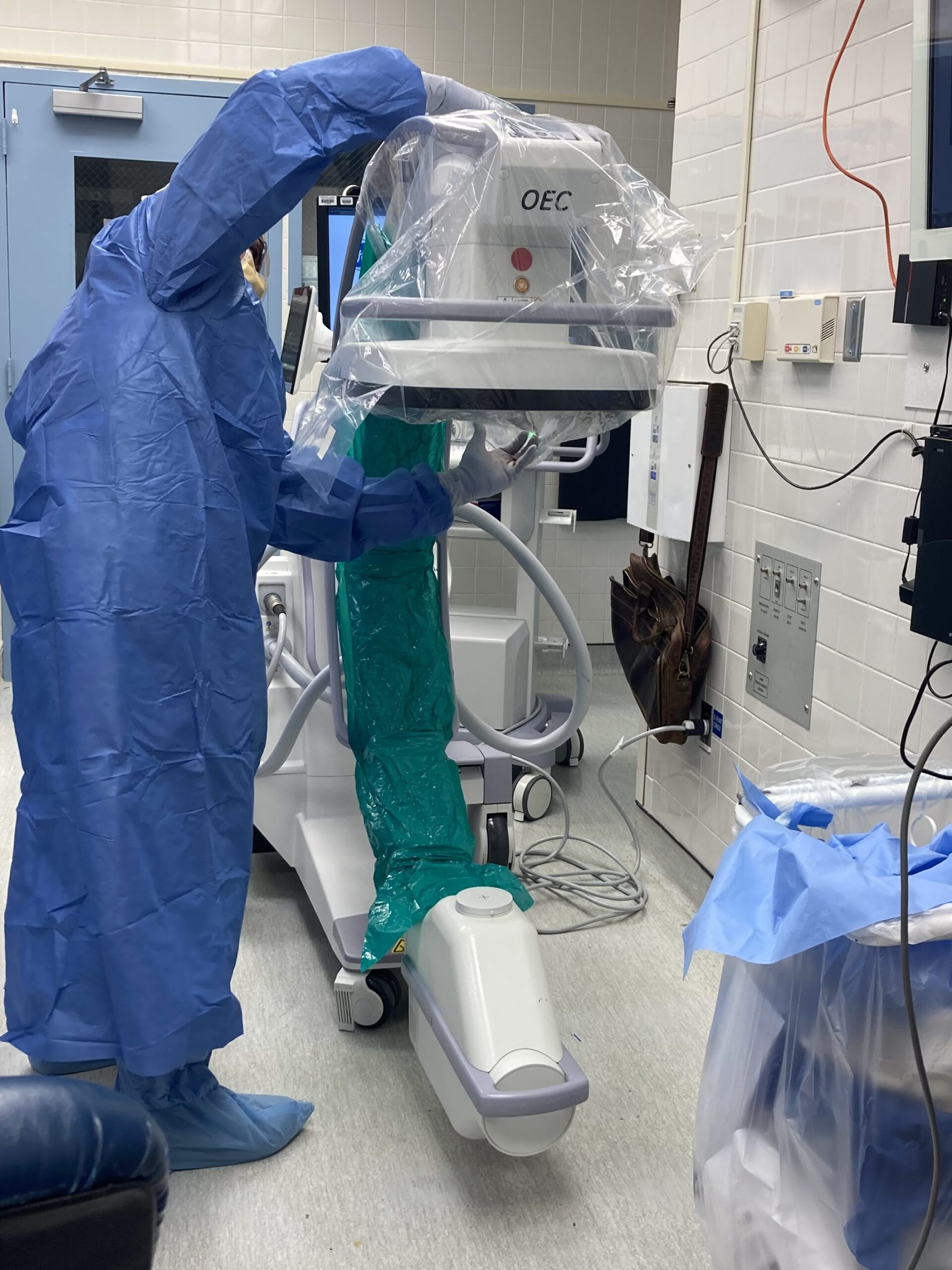
An illustration of how a C-arm is sterile drape for Surgical procedures
2. Cardiac Procedures: In cardiology, C-arms are used for angiography and other cardiac interventions. They enable the visualization of blood vessels, arteries, and the heart in real-time, assisting cardiologists in diagnosing and treating cardiovascular conditions.
3. Pain Management Procedures: C-arms are used in pain management procedures, such as spinal injections, nerve blocks, and other interventions. The real-time imaging allows for precise needle placement, improving the accuracy and effectiveness of the procedure.
4. Gastrointestinal (GI) Procedures: C-arms are utilized in fluoroscopy-guided GI procedures, such as barium studies, swallowing studies, and certain endoscopic procedures. The dynamic imaging helps in evaluating the function and structure of the gastrointestinal tract.
5. Vascular Procedures: C-arms play a crucial role in vascular surgery, including procedures like angiography, embolization, and stent placement. They provide real-time imaging of blood vessels, aiding in navigation and treatment.
6. Trauma and Emergency Care: C-arms are used in trauma settings to assess and treat injuries, especially fractures and dislocations. The real-time imaging capability is valuable in emergency situations to guide quick and accurate decision-making.
Demonstration on how to sterile drape the C-arm
7. Urological Procedures: C-arms are employed in urology for procedures such as kidney stone removal, ureteral stent placement, and other interventions. Real-time imaging helps in navigating the urinary tract during these procedures.
8. Neurosurgery: In certain neurosurgical procedures, C-arms are used for image-guided navigation and localization. This is particularly important in minimally invasive neurosurgical techniques.
9. Critical Care Imaging: C-arms are sometimes used in intensive care units (ICUs) for bedside procedures, such as chest tube insertions, central line placements, and other interventions that benefit from real-time imaging guidance.
O-arm vs C-arm for spine surgery
1. Imaging Technology
O-arm:
Provides 2D fluoroscopic images and 3D volumetric images.
Allows surgeons to see a full 360-degree view of the spine.
It can be used intraoperatively to assess the placement of implants, hardware, and the overall alignment of the spine.
Offers the ability to take a CT-like scan during surgery, which can be critical for complex procedures.
C-arm:
Primarily provides 2D fluoroscopic images.
Typically used for real-time imaging during the procedure.
Less comprehensive in imaging compared to the O-arm but very effective for routine procedures where continuous imaging is required.
2. Use in Surgery
O-arm:
Often used in more complex spinal surgeries, such as deformity corrections or when precise hardware placement is crucial.
It can provide a 3D image at any point during surgery, allowing for immediate correction if something is not positioned correctly.
C-arm:
Commonly used in many types of spinal surgeries, especially where 2D imaging is sufficient.
It’s more portable and easier to use for surgeries where less detailed imaging is required.
3. Precision and Accuracy
O-arm:
Higher accuracy due to its 3D imaging capability, which can be particularly useful in delicate and complex surgeries.
It can reduce the need for post-operative imaging, as it provides detailed information during the procedure itself.
C-arm:
Provides excellent real-time imaging, which is highly useful for monitoring progress during surgery.
While accurate, it doesn’t offer the same level of precision as the O-arm in terms of 3D imaging.
4. Radiation Exposure
O-arm:
Typically, it might expose the patient to more radiation compared to a C-arm, especially during 3D imaging.
However, it may reduce overall exposure by minimizing the need for additional post-operative CT scans.
C-arm:
Generally exposes the patient to lower levels of radiation, especially if only 2D imaging is required.
Continuous fluoroscopy during surgery can increase exposure, but it is usually lower than the cumulative exposure from multiple O-arm scans.
5. Surgeon’s Preference and Experience
O-arm:
Surgeons performing complex spine surgeries often prefer the O-arm for its detailed imaging and accuracy.
C-arm:
Preferred for more routine surgeries or when continuous real-time imaging is necessary.
Conclusion
O-arm: Best for complex surgeries requiring high precision, with the advantage of 3D imaging but higher cost and potentially more radiation exposure.
C-arm: Ideal for routine procedures requiring real-time 2D imaging, more cost-effective, and lower radiation exposure.
The choice between an O-arm and a C-arm depends on the complexity of the surgery, the surgeon’s preference, and the specific needs of the patient.
key safety protocols to follow when using the O-arm and C-arm imaging devices
When using O-arm and C-arm imaging devices in medical procedures, it is essential to follow strict safety protocols to minimize the potential risks associated with ionizing radiation, ensure patient and staff safety, and maintain the overall effectiveness of the imaging equipment.
Training and Certification:
Ensure that all personnel operating or assisting with O-arm and C-arm procedures are adequately trained and certified in radiation safety and the specific use of the imaging equipment.
Radiation Protection Gear:
Use appropriate personal protective equipment (PPE), including lead aprons, thyroid shields, lead gloves, and radiation badges, to minimize radiation exposure to medical staff.
Distance and Shielding:
Maintain a safe distance from the radiation source whenever possible. Use protective shielding, such as lead barriers, to minimize scattered radiation in the operating room.
Time Minimization:
Keep the fluoroscopy or imaging time as short as necessary to achieve the required diagnostic information. Minimize unnecessary exposure to radiation for both patients and medical staff.
Collimation:
Collimate the X-ray beam to the smallest area necessary for imaging. This helps focus the radiation on the specific area of interest and reduces exposure to surrounding tissues.
Regularly calibrate the imaging devices and perform quality assurance checks to ensure accurate and reliable imaging.
Collaboration with Radiology:
Collaborate with radiology professionals to ensure that imaging protocols are optimized for the specific procedure and patient characteristics.
Emergency Shutdown Procedures:
Familiarize all personnel with emergency shutdown procedures in case of equipment malfunction or unexpected situations.
Documentation:
Maintain accurate documentation of radiation exposure levels, imaging parameters, and patient information for each procedure.
Regular Maintenance:
Schedule and perform routine maintenance on the imaging devices to ensure their proper functioning and compliance with safety standards.
Infection Control protocols
Sterilization:
Adhere to proper sterilization protocols for any equipment that comes into contact with the patient or the operating room environment.
Barrier Protection:
Use sterile barriers and covers for imaging equipment to prevent contamination during surgery.
Patient Safety Protocols when using O-Arm and C-Arm
Patient Positioning:
Ensure proper patient positioning to optimize imaging and reduce the need for repeated exposures.
Pregnancy Screening:
Screen female patients of childbearing age for pregnancy before using the imaging devices. If pregnancy is suspected, consider alternative imaging modalities or take additional precautions.
Pediatric Imaging Considerations:
Adjust imaging parameters for pediatric patients to reduce radiation dose. Follow the “As Low As Reasonably Achievable” (ALARA) principle for pediatric imaging
C-Arm Medical Imaging Device and Its History
C-arm is a medical imaging device that provides real-time X-ray images. It consists of a C-shaped arm that houses an X-ray source on one end and a detector on the other. This design allows the C-arm to be maneuvered around a patient to capture X-ray images from various angles without the need for repositioning the patient.
The C-arm was first introduced in 1955 by Dr. Robert S. Ledley, an American physicist and engineer. Dr. Ledley’s invention revolutionized the field of medical imaging by allowing physicians to obtain live X-ray images during surgical procedures, which greatly improved the accuracy and safety of surgeries. Since then, C-arms have become a crucial tool in various medical specialties, including orthopedics, cardiology, and interventional radiology.
Recognizing the C shape on the C-Arm
C-ARM imaging
C-arm devices are commonly used in surgical and interventional procedures where real-time imaging is crucial for guiding the placement of instruments or monitoring the progress of a procedure. They are frequently used in orthopedic surgery, vascular procedures, cardiac catheterization, and other interventions.
The “C” in C-arm comes from the shape of the arm, which is bent like the letter “C”. This allows the device to be easily positioned around a patient during surgery or other medical procedures.
2 main types of C-arms
1. Fluoroscopy C-arm: This type provides continuous X-ray imaging in real-time, which is particularly useful for procedures that require dynamic imaging, such as cardiac catheterization or joint injections.
2. Digital Subtraction Angiography (DSA) C-arm: In addition to real-time fluoroscopy, DSA C-arms have the capability to perform digital subtraction angiography. This technique involves taking a series of X-ray images before and after the injection of a contrast agent. The pre-injection images are digitally subtracted from the post-injection images, highlighting the blood vessels and improving visibility.
C-arm devices play a crucial role in modern medicine, allowing for safer and more precise surgical and interventional procedures. They are used in a wide range of medical specialties and have become an indispensable tool in many healthcare settings.
Recognizing the O that give the O-Arm its name
Disadvantages/Limitation of the C-Arm
Radiation Exposure: One of the most significant limitations is the potential for radiation exposure to both patients and healthcare providers. Prolonged or repeated exposure can be harmful, and measures must be taken to minimize this risk.
Image Quality: Compared to traditional fixed X-ray machines, C-arms may have slightly lower image quality. This is because they are designed for real-time imaging during procedures, rather than high-resolution diagnostic imaging.
Limited Field of View: The field of view (FOV) of a C-arm is generally smaller compared to conventional X-ray machines. This can be a limitation when imaging larger or complex anatomical structures.
Limited Depth Perception: It can be challenging to get a clear understanding of depth and spatial relationships in the images produced by a C-arm. This can make it more challenging to accurately position devices or implants during surgery.
Mobility Constraints: While C-arms are designed to be mobile, they may still have limitations in terms of maneuverability, especially in small or crowded operating rooms. This can sometimes limit the angles and positions from which images can be obtained.
Size and Weight: Some C-arms can be large and heavy, which may limit their use in certain settings or require special accommodations for transportation and storage.
Learning Curve: Operating a C-arm requires specialized training, and not all medical personnel may be familiar with its use. This can lead to a learning curve, particularly for less experienced staff.
Interference with Other Equipment: The use of a C-arm in close proximity to other electronic medical devices can sometimes lead to interference or artifacts in the images.
Limited Soft Tissue Differentiation: Like conventional X-rays, C-arms are better at visualizing dense tissues like bones. Soft tissues, such as organs or muscles, may not be as clearly visualized.
Incompatibility with Certain Procedures: While C-arms are versatile, there may be specific procedures or imaging needs for which they are not well-suited. In such cases, alternative imaging modalities may be required.
Infection Control: C-arms can be difficult to clean thoroughly due to their complex design, which can pose challenges for infection control in a clinical setting.
8 key features of the O-Arm vs C-Arm
O-ARM IMAGINE DEVICE
C-ARM IMAGING DEVICE
1. The O-arm is a specialized imaging system designed in a ring shape, providing a 360-degree scan around the patient. It creates high-resolution 3D images
1. The C-arm is named for its C-shaped arm, which can be maneuvered around a patient. It provides real-time X-ray imaging from various angles without repositioning the patient.
2. It primarily focuses on high-resolution 3D imaging, which is particularly useful for complex surgeries like spinal procedures and orthopedic surgeries. It can also provide 2D fluoroscopy.
2. C-arms primarily provide real-time X-ray imaging (fluoroscopy) but may also have capabilities for digital subtraction angiography (DSA). They are versatile and used in a wide range of procedures including orthopedic, vascular, cardiac, and other interventions.
3. Applications: Spinal surgery (e.g., spinal fusion, pedicle screw placement) Orthopedic procedures (e.g., joint replacement, fracture repair) Neurosurgery Trauma surgery
4. Provides high-resolution 3D images, which are particularly beneficial for procedures that require precise anatomical visualization.
4. Provides real-time X-ray imaging, which is crucial for procedures that require immediate feedback and dynamic guidance
5. Typically more expensive due to its advanced 3D imaging capabilities.
5. Generally more cost-effective compared to O-arm systems.
6. Requires a dedicated space due to its ring-shaped design.
6. More compact and versatile, making it easier to maneuver and fit into a variety of surgical settings.
7. Requires specialized training for optimal use, particularly for 3D imaging.
7. More commonly used and thus, more familiar to a broader range of medical professionals.
8. Typically involves lower radiation exposure for patients and surgical staff due to its advanced imaging techniques.
8. Involves a higher radiation dose compared to O-arm, especially during fluoroscopy.
C-Arm: Price Range: C-arms can vary widely in price depending on factors like brand, model, features, and whether they are new or refurbished. Generally, you can expect a new C-arm to cost anywhere from $100,000 to over $1 million. Refurbished units may be available at a lower cost, typically ranging from $40,000 to $500,000. O-Arm: Price Range: O-arms, also known as intraoperative CT scanners, tend to be significantly more expensive than C-arms due to the advanced imaging capabilities they offer. A new O-arm system can cost between $1 million to $3 million or even more, depending on the specific model and configuration. Refurbished O-arms may also be available at a lower cost, but they are still a significant investment.
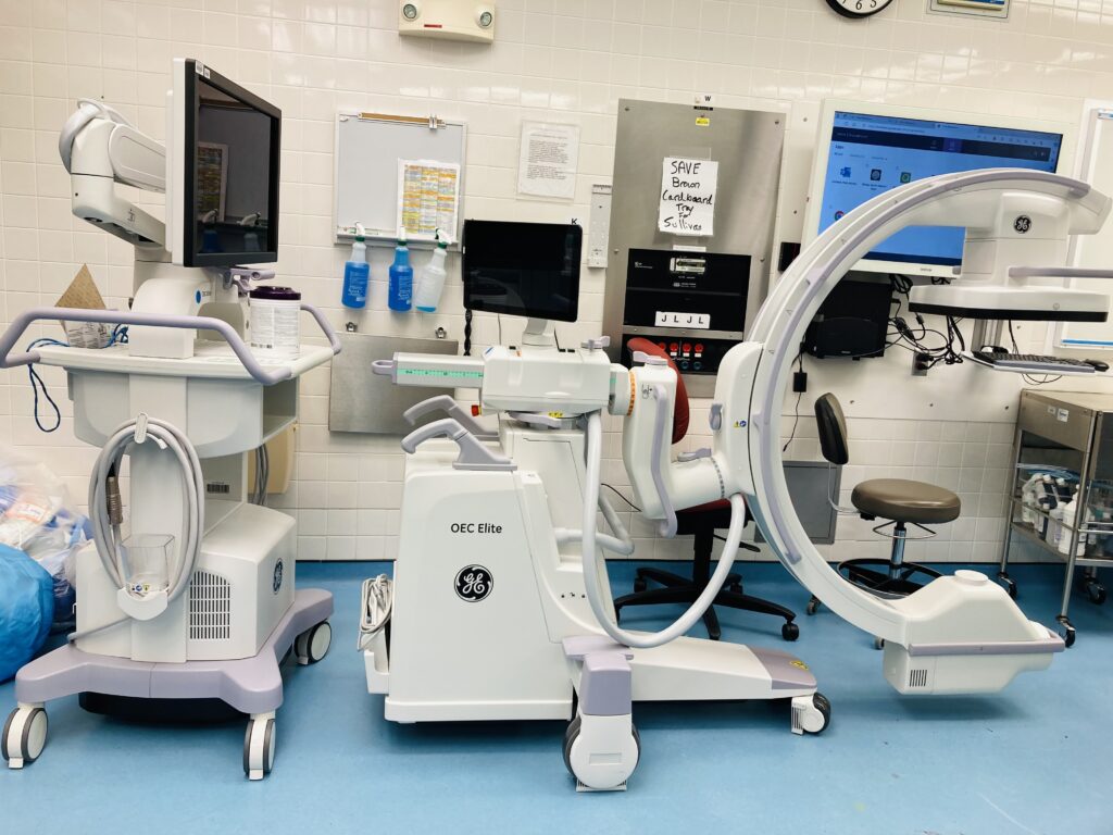
4 key Advancements of the C-Arm
OEC elite C-Arm imaging device
1. Flat-Panel Detectors (FPDs): Traditional C-arms utilized image intensifiers and analog cameras, but modern systems employ FPDs. These detectors offer higher image resolution, improved contrast, and wider dynamic range compared to their predecessors.
They also enable real-time imaging with minimal distortion, enhancing visualization during procedures.
2. Dose Reduction Techniques: Concerns about radiation exposure have driven innovations in dose reduction techniques for C-arm imaging. Manufacturers have introduced technologies such as pulsed fluoroscopy, automatic exposure control, and advanced image processing algorithms to minimize radiation dose while maintaining image quality.
These techniques are crucial for ensuring patient safety and reducing the long-term risks associated with radiation exposure.
3. Augmented Reality (AR) Guidance Systems: AR guidance systems integrate virtual information directly into the surgical field, providing surgeons with real-time navigation assistance during procedures.
By overlaying anatomical structures, preoperative imaging data, and critical procedural information onto the live fluoroscopic images, AR systems enhance surgical precision and reduce the likelihood of errors. Surgeons can visualize complex anatomy, plan trajectories, and accurately place implants with greater confidence and efficiency.
4. Robotic-Assisted Imaging: Robotic-assisted imaging systems combine the capabilities of C-arm technology with robotic manipulation, offering enhanced flexibility, accuracy, and control during surgical procedures.
These systems utilize robotic arms to precisely position the C-arm in desired orientations, enabling complex imaging trajectories that may be difficult to achieve manually. Robotic-assisted imaging enhances workflow efficiency, reduces radiation exposure for surgical staff, and improves patient outcomes by facilitating minimally invasive procedures with greater precision.
Latest O-Arm And C-Arm Advancement
Commercial Launch of O‑arm 4.3 Software Medtronic officially introduced the O‑arm 4.3 software—including 3D Long Scan, Spine Smart Dose, and Medtronic Implant Resolution (MIR)—on September 25, 2024, during the North American Spine Society (NASS) Annual Meeting
Regulatory Milestone Additionally, the software update for the O‑arm O2 system (version 4.3.0), encompassing the same key features, was documented in an FDA 510(k) submission prepared on February 16, 2024
O-Arm Highlights (Medtronic)
O‑arm™ 4.3 (Software Update): Now available in both Europe and other regions, this enhancement introduces key features:
3D Long Scan — Expands imaging coverage up to ~43.8 cm, giving surgeons a wider anatomical view.
Spine Smart Dose (AI‑enhanced) — Cuts radiation dose by approximately 30% while preserving image quality.
Medtronic Implant Resolution (MIR) — Enhances visualization around implants, aiding evaluation of screw placements.
Modern Integration & Safety: The O‑ARM system supports multiple fields of view, low-dose modes, and real-time 2D/3D imaging. It’s optimized for AI and VR integration, streamlining intraoperative scanning and reducing the need for separate pre- or post-op scans.
C-Arm Advances
Siemens Healthineers – CIARTIC Move
A self-driving mobile C‑arm with FDA 510(k) clearance.
Features include:
Motorized chassis with holonomic (omnidirectional) wheels.
Stores up to 12 preset positions with imaging parameters.
Remote-controlled adjustments—even from within the sterile field.
Active collision-sensing for improved safety.
Reduces reliance on tech staff and operator strain.
Philips – Zenition Series
Zenition 30 (Mobile C‑arm):
FDA 510(k) cleared (April 2024).
Offers enhanced image quality, personalization, and dose efficiency.
Zenition 90 Motorized:
Also FDA-cleared (mid‑2024).
High-power (25 kW) motorization and intuitive table-side control to meet complex vascular and OR workflow demands.
Forward-Looking Trends
Flat-Panel Detectors: Philips’ Veradius C‑arm now uses thin flat detectors for improved image resolution, compact footprint, and up to 75% lower radiation via pulsed fluoroscopy.
AI & 3D Enhancements: Mobile C‑arms are increasingly featuring 3D imaging, real-time guidance, and AI-driven workflows—boosting efficiency, reducing radiation, and expanding intraoperative capabilities